What is AI learning to see during liver mobilization?
- Publication state
- PublishedPublished after author review.
- Evidence basis
- 2Connected public source records listed below.
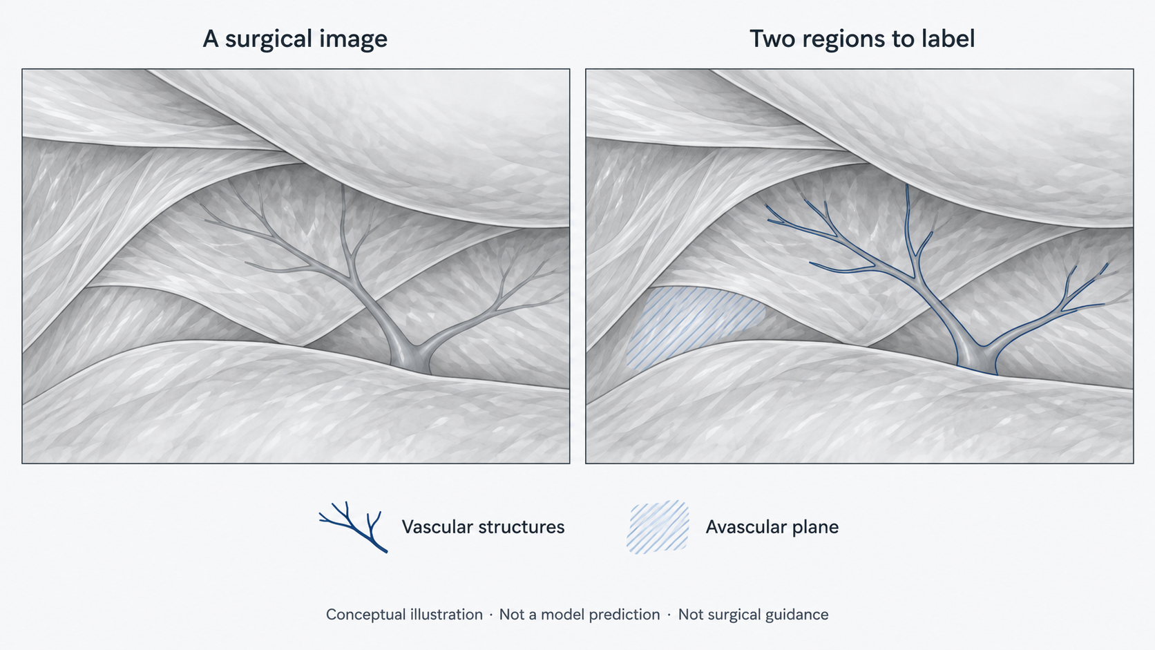
In this study, AI was trained to label vascular structures and the avascular plane in images of laparoscopic donor liver surgery. The output is an anatomical annotation, not a guarantee that dissection is safe.
In the procedures studied, the right liver was mobilized from surrounding attachments before the liver tissue was divided. At this step, a useful visual aid would need to distinguish vessels from the plane being exposed—not simply recognize that a liver is on screen.
The 2025 study coauthored by Namkee Oh addressed this narrower task. Experienced surgical staff outlined the two categories in recorded surgical images, and surgeons reviewed those labels. The model then learned to predict the corresponding regions, a process called semantic segmentation. Its evaluation used 48 surgical videos from three hospitals, including an external test set.
One way to understand this design is as a translation problem: which part of a surgeon’s visual judgment can be made explicit enough to label, compare, and test? Choosing the labels is therefore not just preparation for an AI model. It defines the clinical question the model can—and cannot—answer.
The important boundary is that an “avascular plane” label represents an interpretation of an image. It does not certify every action taken in that region. This retrospective study did not test whether using the system reduced bleeding or vascular injury. It offers an early step toward visual assistance, while clinical benefit still requires prospective evaluation.
AI는 간을 박리하는 화면에서 무엇을 배우는가?
이 연구의 AI는 복강경 간 공여자 수술 영상에서 혈관 구조와 무혈관 박리면을 표시하도록 학습했습니다. 그 결과는 해부학적 영역의 표시이지, 그곳을 박리해도 안전하다는 보증은 아닙니다.
이 연구가 다룬 수술에서는 간 실질을 절단하기 전에 오른쪽 간을 주변 부착 조직으로부터 분리하는 가동화 과정을 거쳤습니다. 이때 유용한 시각 보조 도구가 되려면 화면에 간이 있다는 사실만 알아보는 데 그치지 않고, 노출되는 박리면과 혈관을 구분할 수 있어야 합니다.
오남기가 공동저자로 참여한 2025년 연구는 이처럼 범위를 좁힌 과제를 다뤘습니다. 숙련된 수술 인력이 녹화 영상에 두 범주의 영역을 표시하고, 외과의가 이를 검토했습니다. 모델은 이 표시를 학습해 해당 영역을 예측했습니다. 이를 의미론적 분할, 즉 semantic segmentation이라고 합니다. 평가는 외부 검증 자료를 포함해 세 병원의 수술 영상 48건을 이용했습니다.
이 설계를 이해하는 한 가지 관점은 ‘번역’입니다. 외과의가 눈으로 판단하는 내용 중 무엇을 명시적인 영역으로 표시하고, 비교하며, 검증할 수 있을까요? 무엇에 이름을 붙일지 정하는 일은 단순한 AI 학습 준비가 아닙니다. 모델이 답할 수 있는 임상 질문과 답할 수 없는 질문을 정하는 작업이기도 합니다.
여기에는 중요한 경계가 있습니다. ‘무혈관 박리면’이라는 표시는 영상에 대한 해석이며, 그 영역에서 이루어지는 모든 행위의 안전성을 보증하지 않습니다. 이 후향적 연구는 시스템 사용으로 출혈이나 혈관 손상이 줄어드는지 검증하지 않았습니다. 시각적 보조를 향한 초기 단계이며, 임상적 유익은 앞으로 전향적으로 평가해야 합니다.