Why compare CT liver volume with actual graft weight?
- Publication state
- PublishedPublished after author review.
- Evidence basis
- 3Connected public source records listed below.
Because reproducing another reading of a CT scan is not the same as connecting an image-derived estimate to the graft used in transplantation. Comparing volume with actual graft weight tests that clinically relevant relationship.
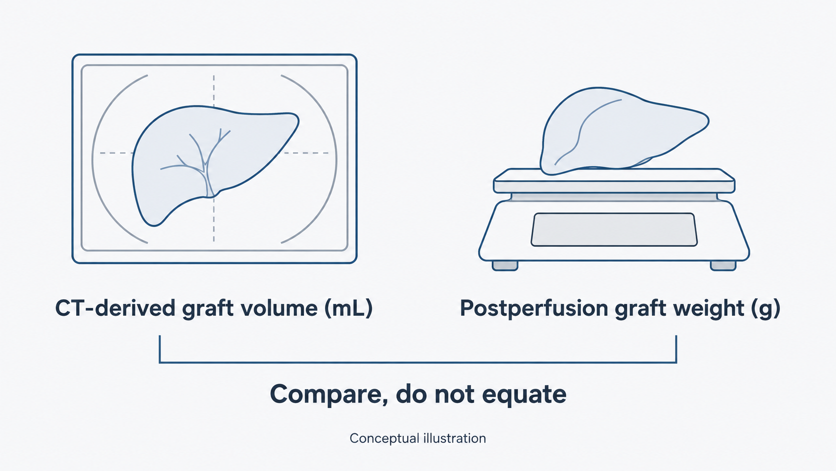
In living donor liver transplantation, a preoperative scan provides a view of the planned graft while it is still inside the donor. The graft can then be weighed after procurement. Volume and weight describe different properties, in different units; one should not simply be relabeled as the other.
A 2024 study by Namkee Oh and colleagues evaluated deep-learning-assisted CT angiography using both surgeon-derived volumes and actual graft weights. For the latter comparison, nine donors were analyzed. AI-derived volume was associated with graft weight, with a reported R² of 0.76. Weight was measured after procurement and perfusion.
The important design choice was to look beyond agreement between image measurements. The study also asked whether the imaging estimate tracked a measurement obtained from the graft itself.
This does not make R² a percentage accuracy score. Nor does correlation alone tell a surgeon how many grams an estimate could be wrong for an individual donor. Those are separate questions for evaluating a prediction tool.
The editorial takeaway is straightforward: assess a surgical imaging tool against the clinical question it is intended to inform, not only against another image annotation.
This was a small, single-center analysis. The findings warrant further validation; they do not establish a universal volume-to-weight conversion or improved donor or recipient outcomes.
CT로 측정한 간 부피를 실제 이식편 무게와 비교하는 이유는 무엇일까요?
CT 영상의 다른 측정 결과를 재현하는 것과, 영상에서 얻은 추정치를 실제로 이식하는 간과 연결하는 것은 다른 문제이기 때문입니다. 부피와 실제 이식편 무게를 비교하면 임상적으로 의미 있는 이 관계를 평가할 수 있습니다.
생체간이식에서 수술 전 영상은 이식할 간이 아직 공여자 몸 안에 있는 상태를 보여줍니다. 간을 적출한 뒤에는 그 무게를 측정할 수 있습니다. 부피와 무게는 서로 다른 속성이며 단위도 다릅니다. 한쪽 값을 그대로 다른 쪽 값으로 바꾸어 불러서는 안 됩니다.
오남기와 공동연구진의 2024년 연구는 딥러닝 기반 CT 혈관조영 영상 분석을 외과의의 부피 측정값 및 실제 이식편 무게와 비교했습니다. 무게와의 상관분석에는 공여자 9명이 포함됐습니다. AI로 추정한 부피와 이식편 무게의 관계에 대해 논문은 R²=0.76을 보고했습니다. 무게는 적출·관류 후 측정했습니다.
주목할 설계상의 선택은 영상 측정값 사이의 일치도만 확인하는 데 그치지 않았다는 점입니다. 영상에서 얻은 추정치가 이식편 자체에서 측정한 값과도 관련되는지 살펴봤습니다.
그렇다고 R²가 백분율 정확도를 뜻하는 것은 아닙니다. 상관관계만으로는 개별 공여자에서 추정치가 몇 그램까지 틀릴 수 있는지도 알 수 없습니다. 이는 예측 도구를 평가할 때 별도로 확인해야 할 질문입니다.
이 글에서 제안하는 해석은 간단합니다. 수술 영상 도구는 다른 영상 주석과 얼마나 비슷한지만이 아니라, 그 도구가 도움을 주려는 임상적 질문에 비추어 평가해야 합니다.
이번 분석은 단일기관의 소규모 비교였습니다. 추가 검증이 필요하며, 보편적으로 적용할 부피–무게 환산법이나 공여자·수혜자의 임상 결과 개선을 입증한 것은 아닙니다.
Connected public evidence
Comprehensive deep learning-based assessment of living liver donor CT angiography: from vascular segmentation to volumetric analysis · PMID: 38869975
Source article · International Journal of Surgery (2024) · DOI: 10.1097/JS9.0000000000001829
Open-access full text · Study methods and graft-weight comparison · PMCID: PMC11487025